NATIONAL REFERENCE LABORATORY (NRL) for Diagnosis of Parasitic Diseases
Working hours: Monday to Friday from 7:30 to14:30
Head: Associate Professor Nina Dimitrova Tsvetkova, PhD
E-mail: This email address is being protected from spambots. You need JavaScript enabled to view it.
Phone: +359 2 843 80 02; +359 2 94 46 999/ 316; 329; 311
Fax: +359 2 843 80 02
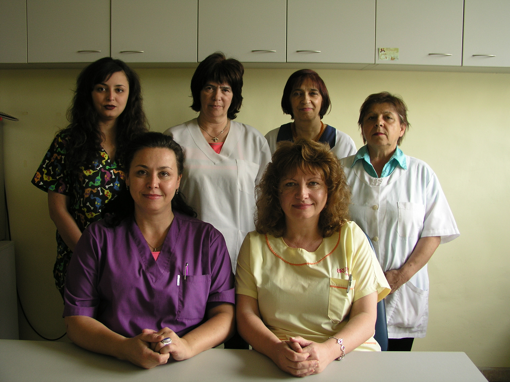
Laboratory staff:
Raina Borisova, MD
Aleksandra Ivanova, biologist,
Mihaela Vanyova Videnova, biologist
Violeta Chavdarova Yakimova, biologist
Galina Mesdraliiska, technical assistant
Silvia Koleva – laboratory worker
The Ministry of Health (MH) has attributed to the laboratory the official status of National Reference Laboratory for the Diagnosis of Parasitic Diseases (NRLDPD). The laboratory is in charge of the organization and implementation of a National System for External Quality Assurance (EQA) of parasitic laboratory diagnosis carried out by all parasitological laboratories in the country. NRLDPD is included in the German Scheme of Quality Assurance - Instand. The laboratory has obtained accreditation status according to the standard EN ISO/IEC 17025:2001. Reference diagnosis of malaria, toxoplasmosis, leishmaniasis, amebiasis, trichinellosis, echinococcosis, pneumocystosis, toxocarosis is performed.
All parasitological laboratories and clinical departments in the country are provided with technical assistance. NRLDPD is also in help of the MH and Regional Health Inspectorates in cases of outbreakes and epidemics. Epidemiological work and annual analysis of the parasitic morbidity is conducted as well as evaluation of the activities of the parasitological network in the country. The laboratory staff is involved in postgraduate education.

The laboratory carries out routine and reference diagnosis by applying contemporary laboratory methods – morphological, immunological and biomolecular.
1. Intestinal helminthic diseases: Аscariasis, Trichuriasis, Enterobiasis, Тaeniasis, Hymenolepiasis, Strongyloidiasis, Ancylostomiasis, Diphyllobothriasis, Fascioliasis, Dicrocoeliasis, intestinal Schistosomiasis and other rare or tropical helminthic diseases. Material: stool
- Маcroscopic examination and morphological differentiation of intestinal helminths
- Microscopic examination for helminth eggs and larvae:
- Wet mount preparations – with saline and iodine solutions
- Concentration procedures (sedimentation, flotation, formalin - ethyl acetate sedimentation technique)
- Detection and larvae cultivation - Baermann funnel technique, Harada-Mori filter paper technique
- Transparent tape test: Enterobiasis and Taeniasis (T. saginata)
- Immnodiagnostic methods in Fascioliasis (ELISA, IHA, IFA)
2. Intestinal protozoal diseases (Giardiasis, Amebiasis, Balantidiasis, Blastocystis infection, Cryptosporidiosis, Cystoisosporiasis, Cyclosporiasis, Мicrosporidiosis). Materials: stool, duodenal material (Giardiasis), biopsy material:
- Мicroscopic examination for trophozoits, cysts and oocysts:
- Wet mount preparations – with saline and iodine solutions
- Concentration procedure – formalin - ethyl acetate sedimentation technique
- Staining procedures – Giemsa, trichrome, modified Ziehl Neelsen staining, etc.
- Cultivation: Pavlova’s medium (Аmebiasis, Blastocystis infection)
- Immunodiagnostic methods in Amebiasis (ELISA)
- Biomolecular methods: PCR for Giardiasis, Blastocystosis, Cryptosporidiosis
3. Blood and tissue parasitic diseases
Мalaria and Babesiosis. Materials: peripheral (capillary) blood for thick and thin blood films, Rapid Diagnostic Tests and PCR
- Microscopic examination: Staining procedures - Giemsa stain
- Biomolecular methods: PCR for malaria species identification
Visceral leishmaniasis. Materials: bone marrow and serum for immunodiagnostics, as well as biopsy materials from lymph node, liver, and spleen.
- Microscopic examination:
- Staining procedures - Giemsa stain
- Cultivation techniques: cultivation in Novy-MacNeal-Nicolle (NNN) medium
- Immunodiagnostic methods – ELISA, Western blot
- Biomolecular methods – PCR for diagnostics and species identification
Cutaneous and mucocutaneous leishmaniasis: Material: skin lesion biopsy specimens:
- Microscopic examination:
- Staining procedures – Giemsa stain
- Cultivation technique: cultivation in Novy-MacNeal-Nicolle (NNN) medium
Cutaneous amebiasis. Material: bipsy specimen from skin lesion:
- Microscopic examination:
- Staining procedures – trichrome,
- Examination of histological material
- Cultivation technique: cultivation in Pavlova’s medium
Toxoplasmosis. Materials: biopsy specimens - lymph node, spleen, liver, intraocular fluid, placenta, amniotic fluid, as well as cerebrospinal fluid and serum for immunodiagnosis:
- Microscopic examination:
- Staining procedures – Giemsa stain
- Examination of histological materials
- Immunodiagnostics: ELISA (IgG, IgM, IgA antibodies, ELISA – IgG avidity), Western blot
- Intraperitoneal inoculation into mice
- Detection of parasite DNA by PCR
Trypanosomiases (American and African). Materials: venous blood, biopsy material, cerebrospinal fluid
- Microscopic examination:
- Staining procedures – Giemsa stain
- Examination of histological materials
Extraintestinal amebiasis. Material: serum, skin lesion biopsy specimens
- Immunodiagnostics: ELISA, IFA
Naegleria and Acanthamoeba infections. Materials: cerebrospinal fluid, biopsy specimens (brain tissue, skin, cornea) or of corneal scrapings
- Microscopic examination:
- Wet mount preparations
- Staining procedures - trichrome, Heidenhain, Lawless stains
- Examination of histological materials
- Cultivation technique – non-nutrient agar (NNA) plates, PPG medium
- Моlecular methods - PCR
Pneumocystosis. Materials: induced sputum, bronchoalveolar lavage, biopsy specimen
- Microscopic examination:
- Staining procedures – Giemsa, toluidine blue
Еchinococcosis. Materials: serum sample, pathological specimens for detection of present scolexes in the cyst and vitality of the parasitic cyst:
- Microscopic examination:
- Examination for presence of parasitic elements – hooks, scolexes and membranes
- Methylene-blue staining for vitality determination
- Immunodiagnostics: ELISA, Western blot
Toxocariasis (Toxocarosis). Material: serum sample:
- Immunodiagnostics: ELISA, Western blot
Тrichinellosis (Trichinosis). Materials: serum sample, muscle biopsy specimen, suspicious meat products for sanitary examination:
- Microscopic examination:
- Examination for presence of trichinella larvae
- Immunodiagnostics – IHA, ELISA, Western blot
- Molecular analysis and Trichinella genotype determination – Multiplex and Nested PCR
- Sanitary examination for presence of Trichinella larvae in meat products
- Compressive trichinelloscopy (Squash preparation)
- Digestion with artificial gastric juice
Cysticercosis. Materials: serum sample, cerebrospinal fluid (CSF), and biopsy histological specimens
- Microscopic examination:
- Examination of hystological specimens for parasitic elements
- Immunodiagnostics - ELISA
Paragonimiasis. Materials: bronchoalveolar lavage, tracheobronchial secretion, induced sputum, biopsy specimen, stool
- Microscopic examination:
- Wet mount preparations
- Concentration techniques
Filariases. Materials: venous blood, biopsy specimen
- Microscopic examination:
- Staining procedures – Giemsa stain
- Concentration techniques: 2% formalin (Knott's technique)
- Examination of subcutaneous nodes (Onchocerca volvulus and Mansonella streptocerca)
- Rapid diagnostic test
Dirofilariases. Material: biopsy specimen
- Мicroscopic examination:
- Examination of histological specimens
- Маcroscopic examination, after parasite extraction
4. Urogenital parasitic diseases.
Тrichomonasis. Materials: urine, semen, prostate exprimate, vaginal and cervical swab specimens, urethral swab specimen:
- Microscopic examination:
- Wet mount preparations
- Staining procedures – Giemsa stain
- Cultivation techniques: cultivation in TV4 medium
- PCR
Schistosomiasis (urogenital). Materials: 24 hours urine, urine collected in the interval between 11.00 and 15.00 o’clock – after physical exercise, bladder biopsy specimen:
- Microscopic examination:
- Wet mount preparations
- Concentration techniques – sedimentation, formalin - ethyl acetate sedimentation technique
- Rapid diagnostic test – with urine
Members of the laboratory participate in the implementation of the following scientific projects financed by the Scientific Research Fund:
Project title: "Molecular genetic studies on the prevalence of human Pneumocystis pneumonia in Bulgaria", project no. KP-06-N33/18 dated 21.12.2019
Duration: 36 months
Project leader: assoc. prof., PhD, Nina Dimitrova Tsvetkova
Pneumocystosis is an opportunistic infection that in immunocompromised individuals, elderly, and young children can lead to serious complications and even death. Early and timely diagnosis, the conduct and monitoring of etiologic treatment are critical to the outcome of the disease.
The aim of the project is to investigate the prevalence of pneumocystosis in different groups of immunocompromised and immunocompetent individuals, to identify the levels of colonization/carrier of the pathogen among at-risk populations, and to clarify the role of colonized individuals as a possible reservoir for the spread of P. jirovecii infection.
The introduction of molecular genetic methods in the diagnosis of pneumocystosis will have a positive effect in several directions, the more important of which are: a) improving the quality of diagnosis of this disease; b) monitoring of the results of conducted treatment by quantifying the pathogen in the clinical material under study; c) accumulation of new data on the epidemiology of P. jirovecii in Bulgaria - to the best of our knowledge, no such studies have been done in the country so far.
In our country the data on the incidence of pneumocystosis need to be updated, expanded, and deepened. This project proposal will be the first study to cover a broad contingent of different patient risk groups to be tested for Pneumocystis pneumonia and colonization/carrier of the pathogen by a combination of diagnostic methods including advanced highly sensitive molecular genetic methods, which will increase the detectability of infected persons.
The use of PCR techniques to demonstrate colonization/carrier of P. jirovecii in clinically healthy individuals (medical staff, relatives or caregivers) coming into contact with patients diagnosed with Pneumocystis pneumonia will lead to increased pathogen detectability due to the potential of molecular genetic techniques to detect a very small number of microorganisms in the analyzed samples, respectively the clinical materials.
The study on gene coding for dihydropteroate synthase (DHPS), which is the target area in the mechanism of action of sulfones and sulfonamides, by restriction fragment length polymorphism (RFLP) analysis will allow new data on the development/lack of resistance of P. jirovecii strains to be obtained and to evaluate the risk of spreading P. jirovecii strains with DHPS gene mutations. This will result in the accumulation of new original data on the circulating strains in our country.
Research methodology and techniques:
Clinical materials (sputum, tracheal aspirate, bronchoalveolar lavage material, mouthwashes, etc. - the type of material depends on the patient's condition) will be tested with the following methods:
1 Standard staining methods for direct microscopy
a) Azure-eosin-methylene blue staining, Romanowsky-Gimza method,
b) Toluidine blue staining (selective method for P. jirovecii cysts),
c) Gomori Methenamine-silver nitrate staining (for P. jirovecii cysts).
2 Molecular genetic techniques
Genomic DNA extraction.
Real-time PCR for detection of P. jirovecii.
PCR-RFLP for detection of point mutations in dihydropteroate synthetase (DHPS) gene.
- Project title: "Studies on the cellular and humoral immune response in patients with acute and chronic form of toxoplasmosis", KP-06-N43/2 of 27.11.2020.
Duration: 36 months
Project leader: Prof. Iskra Raynova, MD, DSc
- Project title: "Biological and molecular genetic studies on the insecticide resistance of native and invasive mosquito species", KP-06-N41/5 dated 30.11.2020.
Duration: 36 months
Project leader: Assistant Professor Ognyan Mikov, PhD
- Project title: "Phylogenetic and genotypic analysis of Enterobius vermicularis and studies on the influence of the parasite on the local immunity of the intestinal tract and the possibility of bacterial co-infections in infected persons in Bulgaria", KP-06-PN53/4 of 11.11.2021.
Duration: 36 months
Project leader: Assistant Professor Eleonora Kaneva, PhD

 English
 Bulgarian